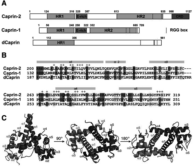
Figure 1. Caprin proteins and structure of the dimerization domain of Caprin-2.

A) Schematic representation of human Caprin-2 and Caprin-1, as well as Drosoplila Caprin (dCaprin). HR1 and HR2: homologous region 1 and 2. CRD: C1q-related domain. The thin red lines indicate the locations of the RGG boxes. B) Sequence of the human Caprin-2 dimerization domain aligned with homologous sequences of Caprin-1 and dCaprin. The secondary structures are indicated above the Caprin-2 sequence. Residues involved in Caprin-2 dimerization are indicate with “+” sign above the sequence. C) Structure of the Caprin-2 homodimer rendered in cartoon mode, viewed from three different angles. The two protomers are coloured red and blue respectively.
