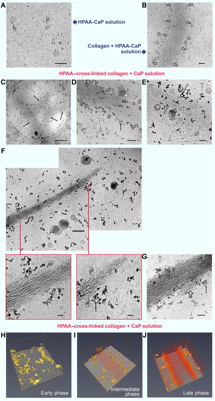
Fig. 2. Cryo-EM of mineralization of bare collagen and HPAA-collagen fibrils.
(A) Grid dipped in freshly prepared HPAA-CaP solution. Scale bar, 50 nm. Prenucleation clusters (open arrowheads) and amorphous calcium phosphate (ACP) droplets (pointers) were randomly distributed within the frozen medium. (B) Grid with bare collagen immersed in HPAA-CaP for 1 hour. Scale bar, 50 nm. Fibrils (open arrows) were not mineralized at this stage. (C to G) HPAA-collagen fibrils were mineralized in unstabilized CaP solution for 1 (C and D), 8 (E), 24 (F), and 72 hours (G). Arrows, prenucleation cluster aggregates; open arrowheads, prenucleation cluster singlets; asterisks, irregular ACP droplets. (C) Circular hole in carbon film (open arrows) shows the concentration of mineral precursors along the periphery of unstained collagen fibrils (arrows). Scale bar, 500 nm. (D) Prenucleation cluster aggregates along the fibril’s surface (arrows) at 1 hour. Scale bar, 100 nm. (E) At 8 hours, fibrils (open arrows) were filled with a slightly more electron-dense material. Scale bar, 50 nm. Prenucleation cluster aggregates were seen in the fibril’s vicinity, while singlets were predominantly located further away. (F) Upper montage of an incompletely mineralized collagen fibril at 24 hours. Scale bar, 100 nm. Bottom left: The intrafibrillar minerals depicted by the pointer [similar to those in (E)] probably represent intrafibrillar ACP that had not yet been transformed into crystalline CaP. Scale bar, 20 nm. Bottom right: Less mineralized part of the fibril with more potentially ACP phases (pointers). Scale bar, 20 nm. (G) Heavily mineralized fibril at 72 hours shows commencement of extrafibrillar mineralization (open arrow). Scale bar, 50 nm. (H to J) 3D rendering of the early (H), middle (I), and late (J) phases of intrafibrillar mineralization of HPAA-collagen showing accumulation of prenucleation cluster aggregates (yellow) along the fibril surface; intrafibrillar minerals are depicted in orange.

