Fig. 3.
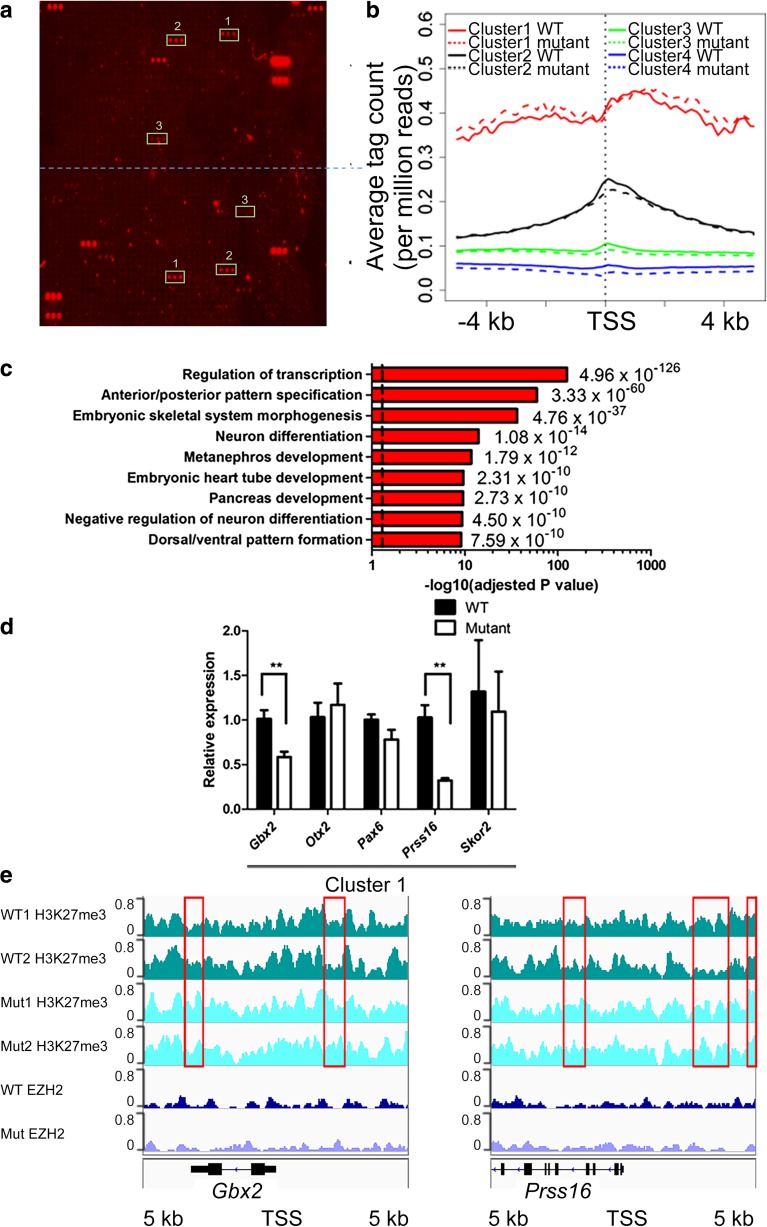
TSPYL2 regulates the level of H3K27me3 in neurons. a Verification of the specificity of H3K27 antibodies for ChIP-seq by hybridization to a histone peptide array. Dotted line divides the sub-arrays. Authentic positive signals appearing in triplets were boxed and peptide identities are 1, H3K27me3 + R36me2; 2, H3K27me3; 3, H4K12ac + K16 ac + H3K27me3. All unmarked triplet peptide signals are from IgG controls to indicate binding of primary antibodies and secondary antibodies. b ChIP-seq results of primary hippocampal neurons at 14 days in culture. H3K27 me3 occupancies at a 10-kb window centering at the TSS were classified into four clusters by k-mean clustering. Metagene analysis shows the increased level of H3K27me3 for cluster1 metagene in Tspyl2 mutant primary neurons. c Selected Gene Ontology of top-enriched biological pathway annotations of cluster 1 genes (DAVID Gene Ontology Bioinformatic Resources, https://david.ncifcrf.gov/home.jsp). Dotted line, P < 0.05 from P value (a modified Fisher’s exact P value) adjusted by Benjamini-Hochberg correction. d RT-qPCR of example marked genes in cluster 1. Transcript level relative to Hprt and the wild-type level are set as 1. n = 4. Error bars represent SEM. **P < 0.01, Student t test. e Genome browser views showing ChIP-seq pileups for Gbx2 and Prss16. Areas of increased H3K27me3 in mutant neurons were boxed. WT: wild-type; Mut: mutant

