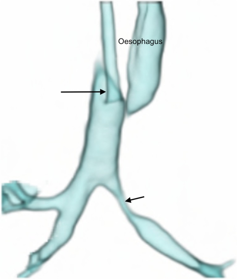
Fig. 2.
Anterior view of a volume-rendered reconstruction from a dynamic 4-D CT study of the airways in a 1-month-old girl with stenosis of the left main bronchus (short arrow). The distal two-thirds of the trachea could be evaluated in this instance because the endotracheal tube was withdrawn to lie with its tip in the upper third of the trachea (long arrow) using the planning scannogram (not shown). Oesophageal air is present and appears on the airway 3-D reconstruction setting. Despite efforts to cut this manually from all four or five phases of the scan, it is often not possible to achieve (as in this case) without encroaching on parts of the airway, due to normal craniocaudal and anteroposterior movement of the airway within the imaged volume, during breathing

