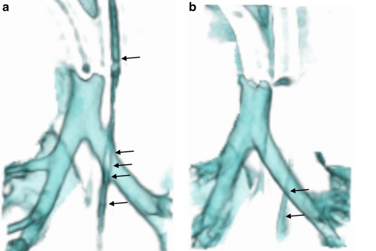
Fig. 3.
Anterior views of 3-D volume-rendered reconstructions of two of the five phases of a dynamic 4-D CT study of the airways in an intubated and ventilated 1-month-old boy with suspected tracheobronchomalacia. a, b Images from two different phases of ventilation demonstrate the nasogastric tube (arrows) crossing the left main bronchus and distorting the outline of the airway during inspiration (a) but not during expiration (b), which could affect assessment of collapsibility of the airway

