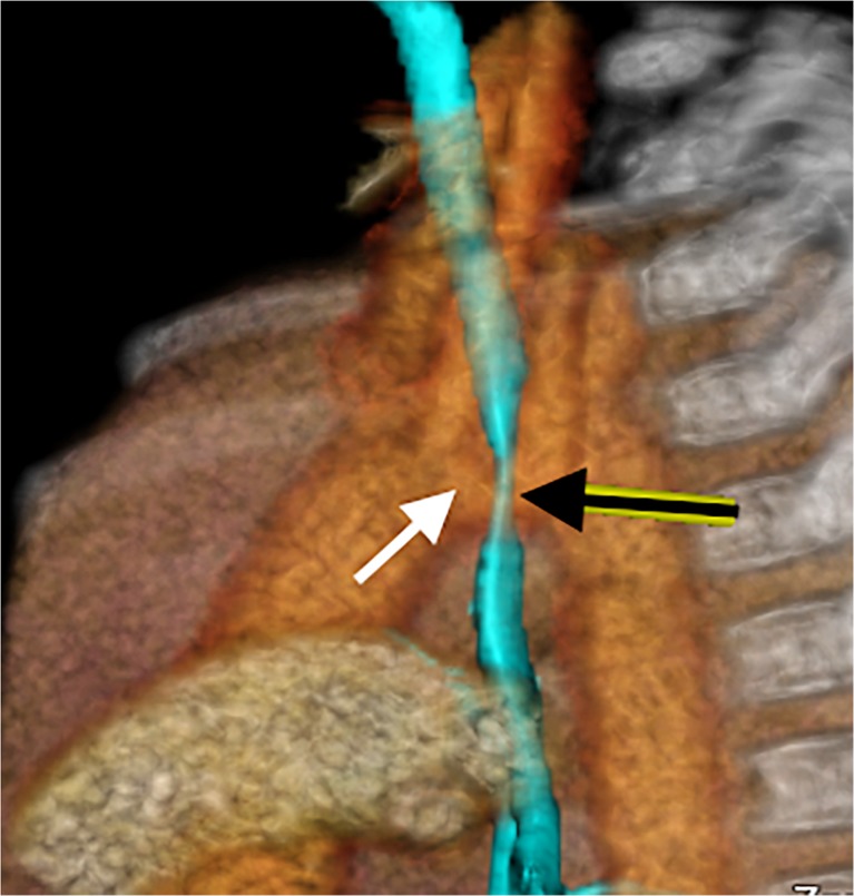
Fig. 9.
A lateral view of a volume-rendered 3-D reformat created from one phase of a dynamic 4-D CT in an 8-month-old boy with right-side arch and innominate impression on the trachea. The relationship of the vascular structures is demonstrated in orange and yellow, with the airways in transparent light blue. Note the close relationship of the left-positioned brachiocephalic trunk (white arrow) and the anterior trachea (black arrow), which on cine mode (Supplementary material 6) demonstrates innominate artery compression syndrome

