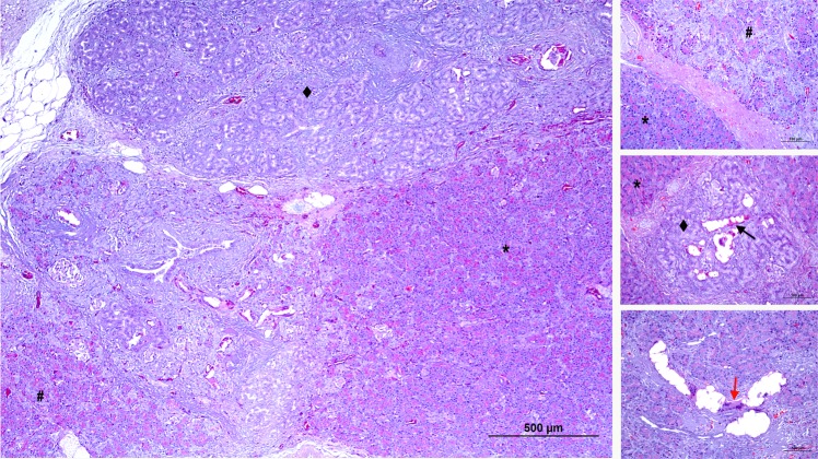
Figure 6.
Microscopic pattern found in the pancreas of the TP-G group (A–C). In this group the loss of the acinar component showed a lobular pattern, with lobules without atrophy (score 0) (*), lobules with irregular and moderate atrophy (score 3) (#), and others with complete disappearance of exocrine pancreatic tissue (score 5) (◆). The glue can be seen inside the ducts of the atrophied lobes (black arrow). In these ducts surrounded by multinucleated cells there is also an obvious foreign body reaction (red arrow).

