Figure 1.
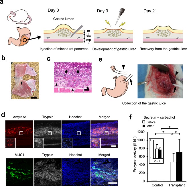
Allogenic transplantation of minced rat pancreas into the gastric submucosal space. (a) Scheme of the transplantation protocol. The suspension of minced pancreas was injected into the gastric submucosal space of the dorsal glandular stomach of rat under laparotomy. Three days after transplantation, the stomach was opened again under laparotomy, and mucosa and muscularis mucosa at the swelling of the transplant site were ablated with electrocautery to develop a gastric ulcer. The ulcer was treated by daily administration of proton pump inhibitor. The various structures of the gastric wall are depicted as – m: mucosa, mm: muscularis mucosa, sm: submucosa, mp: muscularis propria, s: serosa. (b) The stomach specimen. The gastric ulcer scar was observed on the transplant site (arrowhead). Scale bar: 10 mm. (c) Haematoxylin-eosin staining of the transplant site. The transplanted pancreas was observed in the gastric submucosal space (arrowhead) and directly attached to the gastric mucosa without the interference of muscularis mucosa (arrow). Scale bar: 100 μm. (d) Representative images of immunofluorescence staining 3 weeks after transplantation. Top: amylase (red) and trypsin (white). Bottom: MUC1 (green) and trypsin (white). Nuclei were stained with Hoechst 33258 (blue). The transplanted pancreas still expressed the acinar markers amylase and trypsin and the ductal marker MUC1. Scale bar: 100 μm. (e) Gastric juice was collected and the stomach was harvested 21 days after transplantation. Saliva was drained from the oral stump to outside of the abdominal cavity (arrowhead). A 16 G Surflo outer catheter was inserted into the stomach to cleanse the gastric lumen and collect the gastric juice (arrow). (f) Amylase level in the gastric juice. Amylase in the gastric juice was significantly elevated in the transplanted group compared to that in the control group. The administration of carbachol and secretin further increased the amylase level in the transplanted group (n = 5). All data are represented as means ± SD. *p < 0.05.

