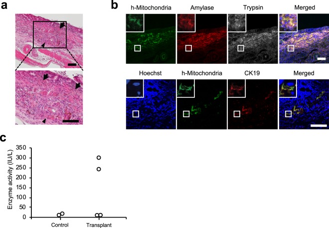
Figure 5.
Implantation of iPSC-derived pancreatic exocrine cells into the gastric submucosal space of rat. (a) Haematoxylin-eosin staining of the implanted cells. The implanted cells were observed in the submucosal space of the stomach (arrowhead), and the implanted pancreas directly attached to the gastric mucosa without the interference of muscularis mucosa (arrow). Scale bar: 100 μm. (b) Representative images of immunofluorescence staining at the implantation site. Top: human mitochondria (green), amylase (red), and trypsin (white). Bottom: human mitochondria (green) and cytokeratin 19 (red). Nuclei were stained with Hoechst 33258 (blue). The implanted iPSC-derived exocrine pancreas still expressed amylase and trypsin. Scale bar: 100 μm. (c) Amylase level in the gastric juice. Amylase in the gastric juice was elevated in two out of four implanted rats, while there was no elevation in the other 2 rats (n = 4). The elevation of amylase was not detected in the gastric juice of the control group (n = 2).

