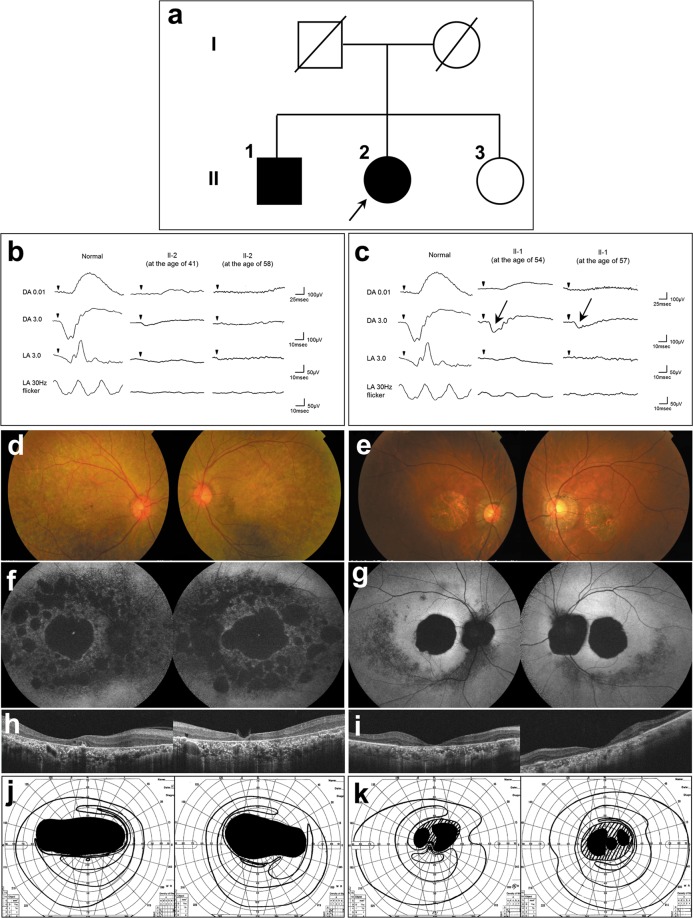
Fig. 1. A pedigree of a Japanese family with CRD and the results of various ophthalmological examinations.
a A pedigree of the Japanese family with CRD. The square boxes and circles denote male and female members, respectively; black symbols indicate affected individuals; and slashed symbols indicate deceased individuals. The proband is indicated with an arrow. b, c Electroretinograms of the proband (b) and her elder brother (c). DA, dark adaptation; LA, light adaptation. For each of the 2 patients, both the results at presentation and the more recent results are shown. The patient’s age at the time of examination is indicated. The scotopic responses in the elder brother’s ERG are indicated with arrows (c). A normal ERG pattern of a healthy individual (from our hospital) was added for comparison. d, e Fundus photographs of the proband (d) and her elder brother (e). f, g Fundus autofluorescence (FAF) of the proband (f) and her elder brother (g). h, i Optical coherence tomography (OCT) of the proband (h) and her elder brother (i). j, k Goldmann visual field of the proband (j) and her elder brother (k)

