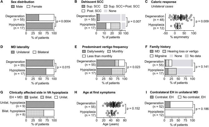
Figure 4.
Clinical differences between and within the two MD endotypes. Data from (A–F,H) are also shown in Table 2, data from (G) and (I) were not part of the initial null hypotheses. Bilat, bilateral; contralat, contralateral; EH, endolymphatic hydrops; ipsilat, ipsilateral; MD, Meniere's disease; post, posterior; SCC, semicircular canal; sup, superior; unilat, unilateral; VA, vestibular aqueduct.

