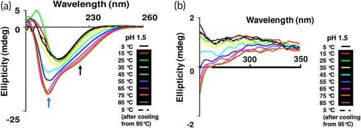
Figure 7.

Combined effect of temperature and pH on the secondary structure of β‐cardiotoxin. Thermal denaturation of β‐cardiotoxin at pH 1.5. The protein was dissolved in MilliQ water (0.5 mg/mL) and pH adjusted to 1.5 with HCl and far‐UV CD spectra (a) and near‐UV CD spectra (b) were recorded using a 0.1 cm path‐length cuvette. Refolding was observed at 5°C after cooling from 85°C, black dotted line. The blue and black arrows indicate the new bands arising at 203 and 222 nm.
