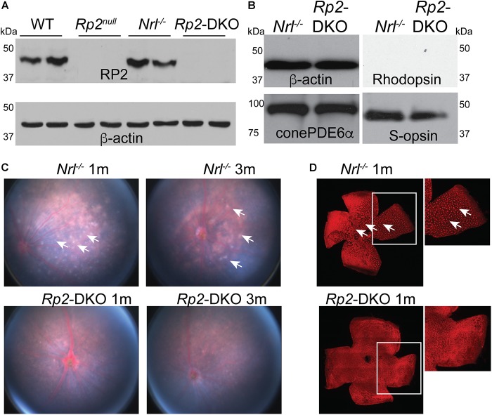
FIGURE 1.
Characterization of the Rp2-DKO mice. (A,B) Retinal extracts (100 μg) from the mice of the indicated genotypes were analyzed by SDS-PAGE and immunoblotting using anti-RP2, rhodopsin, S-opsin, cone PDE6α, and β-actin (loading control) antibodies. Fundus (C) and flat mounted retinal staining (D) of the Nrl-/- and Rp2-DKO mice was performed. Arrows point to the white spots in the fundus photograph that correspond to the whorls and rosettes observed in the PNA (red; peanut agglutinin) stained flat mounted retinas of the Nrl-/- mice. Such spots are undetectable in the Rp2-DKO mouse retinas.

