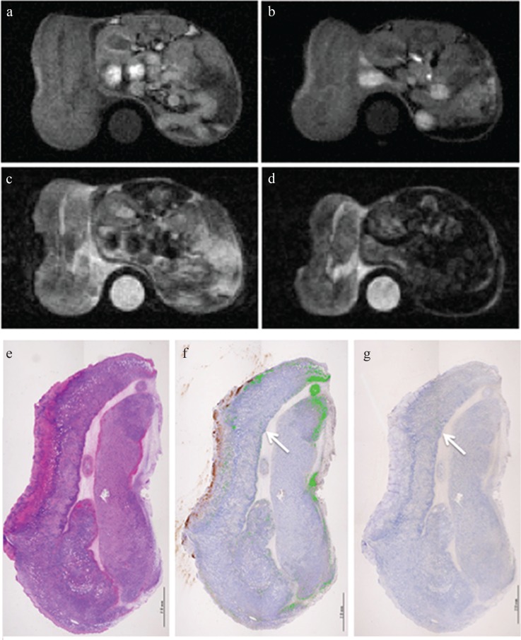
Fig. 6.
MRI and pathology at 24 h after injection of Annexin V-conjugated ultrasmall superparamagnetic iron oxide (V-USPIO) for the mouse treated with anti-tumor drugs (Case 4). Tumor volume change ratio (%) was 62.7%. The tumor signal intensity (SI) on post-T1-weighted MRI (T1WI) (b) was similar or heterogeneously increased compared with that on pre T1WI (a). The post/pre-SI ratio of the whole tumor on T1WI was 1.25. The tumor signal on post T2-weighted MRI (T2WI) (d) was similar or slightly decreased compared with that on pre T2WI (c). The post/pre-SI ratio of the whole tumor on T2WI was 0.91. A round-shaped phantom (water) was also described at the side of the berry in (a–d). (e–g) represent hematoxylin-eosin, Perls DAB and TdT-mediated dUTP nick end labeling (TUNEL) staining, respectively. Perls DAB (%), TUNEL (%) and Necrosis (%) were 23.7, 0.1 and 44.5, respectively. Although the area in which apoptotic cells are observed is limited, the similar distribution of iron and apoptotic cells was observed (arrows). In this case, small foci of hemorrhage were scattered along with a large necrotic area.

