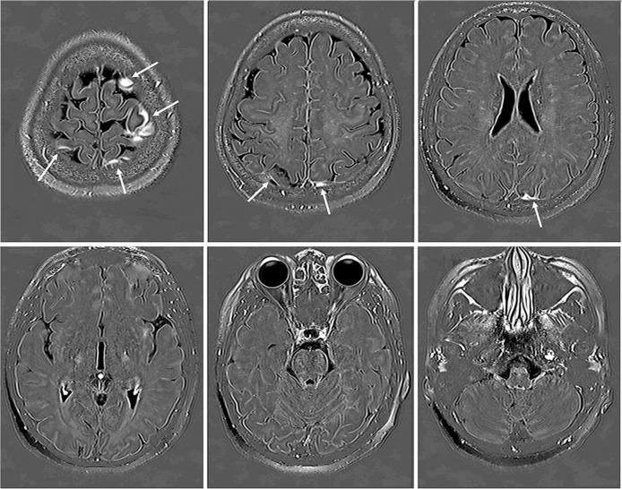
Fig. 2.
A 49-year-old woman with a suspicion of endolymphatic hydrops. In the second part of this study, six out of 256 axial slices obtained at 4 h after intravenous administration of single dose gadolinium based contrast agent (IV-SD-GBCA) were selected at 20 mm increments from the vertex to the skull base for the image evaluation. The cerebrospinal fluid (CSF) around the cortical veins has a high signal intensity on Three dimensional real inversion recovery (3D-real IR)images (arrows).

