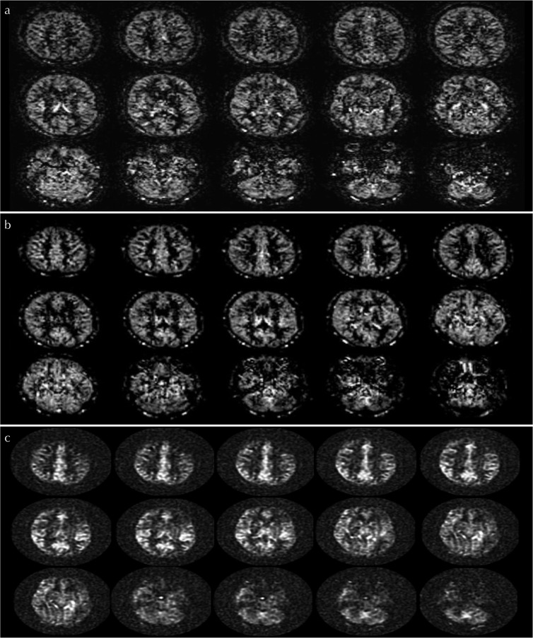
Fig. 4.
Arterial spin-labeling (ASL) perfusion images among 2D-echo planar imaging (EPI), 3D-pseudo steady state (PSS), and 3D spiral-fast-spin echo (FSE) readout. Case examples of ASL perfusion image obtained by 2D-EPI (a), 3D-PSS (b) and 3D-spiral FSE (c) in the same volunteer were respectively presented.

