
Figure 1. Intermittent fasting (IF) ameliorates EAE clinical course and pathology.
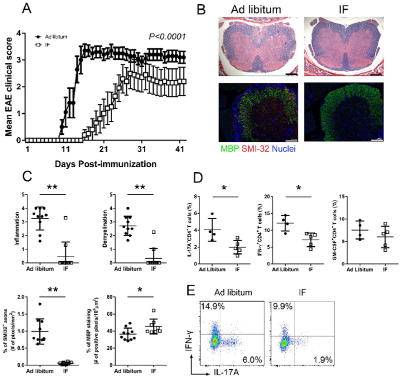
C57BL/6 mice underwent IF or were fed ad libitum (control group) for a month before immunization (n=10/group). (A) EAE clinical course of a representative experiment (dots represent the mean clinical scores for all 10 mice in each group, error bars are SEM.; P<0.0001 by two-way ANOVA). Four EAE experiments were performed with similar results. (B) Spinal cord pathology: the upper panel shows a histological staining (solochrome cyanine) for myelin (in blue) and the lower panel is an immuno-staining for SMI-32+ damaged axons (in red) and MBP (in green). (C) Quantification of inflammation, demyelination (evaluated by histology and MBP staining) and axonal damage (evaluated by SMI-32+ staining) in the spinal cord in the two groups (n=10/group) on day 26 post-immunization. Each dot represents a mouse and the bars are means ± SD. (D) Percentages of CD4+ T cells producing IL-17A, IFN-γ and GM-CSF measured by flow cytometry in lymph nodes draining the immunization site on day 6 post-immunization. Each dot represents a mouse and the bars are means ± SD. This is one of three different experiments performed with similar results (n=4–5/group in each experiment; Table S2 reports the results of all experiments performed). (E) Representative flow-cytometry plots for T cell production of IL-17A and IFN-γ in CD4+ T cells isolated from the draining lymph nodes at day 6 post-immunization in the two groups. * P<0.05; ** P<0.005. All P values were calculated by Mann-Whitney test.