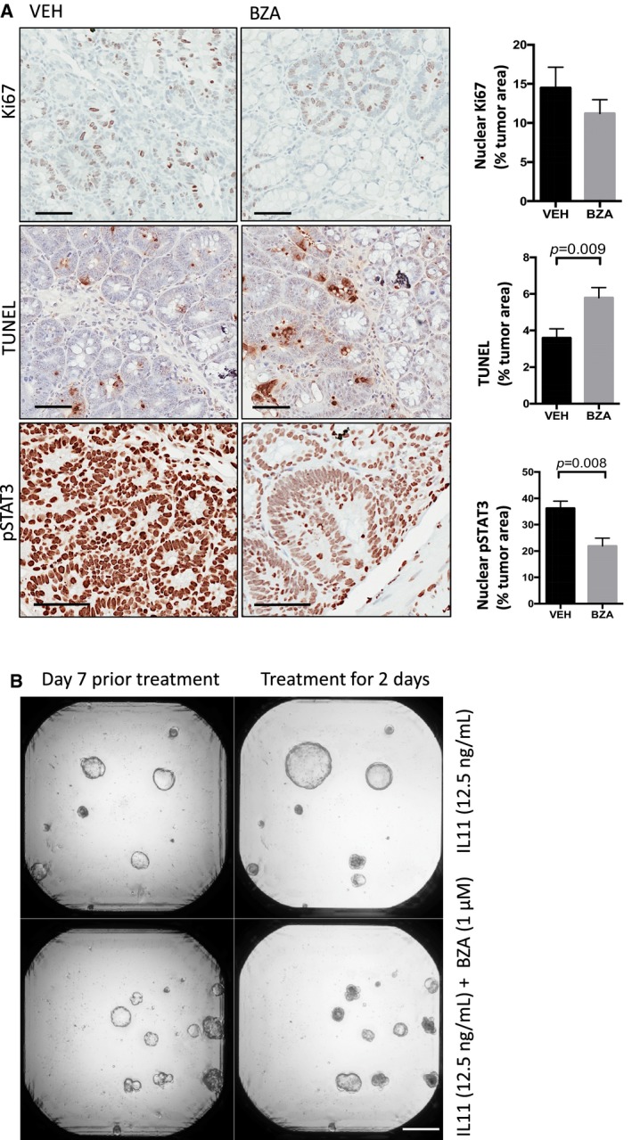
Figure EV3. Bazedoxifene treatment suppresses proliferation and induces apoptosis of colonic tumor epithelium in Cdx2 Cre ERT 2; Apc flox mice.

-
ARepresentative immunohistochemistry staining for proliferative (Ki67 positive) and apoptotic (TUNEL positive) epithelium and those cells with high levels of the transcriptionally active pSTAT3 isoform (3+ intensity score) in tumor sections of mice as treated in Fig 5A. Immunohistochemical staining was quantified by Aperio analysis as outlined in the Materials and Methods section. Scale bars = 100 μm. Data are mean ± SEM, with n = 5 mice per cohort, P‐value derived from unpaired Student's t test.
-
BRepresentative images of colon cancer organoids derived from Apc Min mice grown for 7 days in the presence of IL11 (12.5 ng/ml) alone or in combination with 1 μM bazedoxifene (BZA) for a further 2 days. Scale bar = 500 μm.
