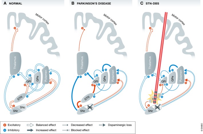
Figure 5. Effects of Parkinson's disease and DBS on basal ganglia networks.

Basal ganglia models: (A) normal state with physiological balanced output to the motor cortex (B) in Parkinson's disease the dopaminergic loss in the SNc leads to widespread excitatory and inhibitory changes. The STN receives fewer inhibitory signals from the GPe and therefore sends greater excitatory signals to the main basal ganglia output structures (GPi and SNr). The thalamus is inhibited and sends fewer excitatory information to the motor cortex resulting in rigidity and bradykinesia (C) with STN‐DBS the nucleus is uncoupled from its afferents. The excitatory output to the GPi and SNr is reduced which in turn send a more balanced signal the motor cortex via the thalamus resulting in reduced rigidity and bradykinesia. [This model is simplified and leaves the hyperdirect pathway from the motor cortex to the STN and the loop between STN and the pedunculopontine nucleus (PPN)]. GPe: globus pallidus pars externus; GPi: globus pallidus pars internus; SNc: substantia nigra pars compacts; SNr: substantia nigra pars reticulata; and STN: subthalamic nucleus.
