Fig. 5.
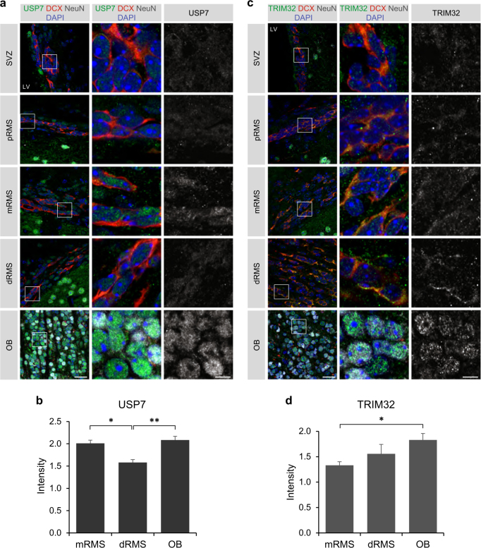
TRIM32 and USP7 show distinct expression patterns in neuroblasts of the distal RMS in vivo. a–c Immunostainings of adult mouse brain sections with the indicated antibodies. Images were taken from the indicated regions of the SVZ-RMS-OB system. The right panels show a higher magnification of the boxed areas. Scale bars = 20 µm; for high magnifications, 5 µm; N = 3. b–d Quantification of the staining intensity for USP7 (b) and TRIM32 (d) in the middle RMS, distal RMS and OB, normalized to the background of each section (mean ± SEM; N = 3; t-test, **p < 0.01; *p < 0.05). pRMS proximal RMS, mRMS middle RMS, dRMS distal RMS

