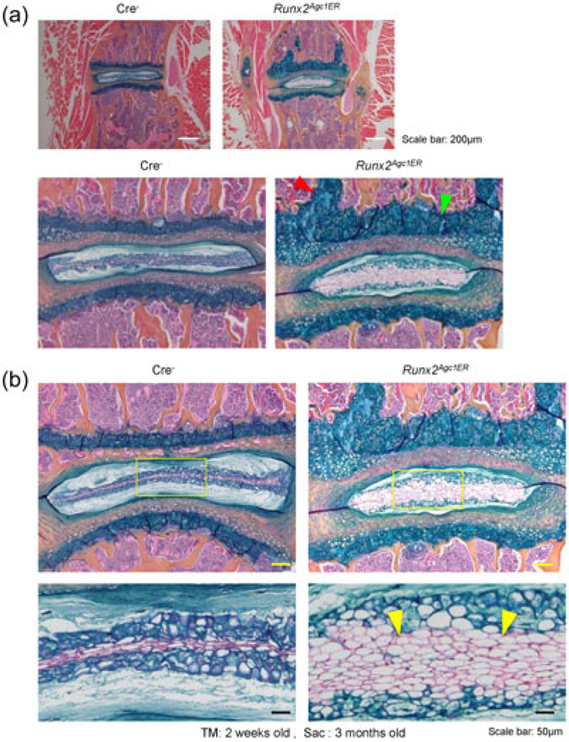
FIGURE 3.

Defects in disc tissues of Runx2Agc1ER KO mice. (a) Results of histologic analysis showed that the growth plate thickness was significantly increased in Runx2Agc1ER KO mice, suggesting that Runx2 inhibits growth plate cartilage growth in normal mice (green arrowhead: increased growth plate cartilage thickness; red arrowhead: the expanded growth plate tissue was protruded to the vertebral body). (b) Increased numbers of notochordal cells in NP were observed in Runx2Agc1ER KO mice (yellow arrowheads: notochordal cells). KO: knockout; NP: nucleus pulposus; Runx2: runt-related transcription factor 2 [Color figure can be viewed at wileyonlinelibrary.com]
