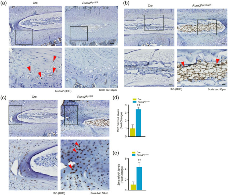
FIGURE 4.

Changes in Ihh signaling in disc tissues of Runx2Agc1ER KO mice. Two-week-old Cre− control and Runx2Agc1ER KO mice were treated with tamoxifen and immunohistochemical (IHC) assay was performed using disc tissues of 3-month-old Cre− and Runx2 KO mice. (a) Results showed that Runx2 expression (red arrowheads indicate the Runx2-positive cells in Cre− mice) was significantly reduced in the annulus fibrosus (AF) cells of Runx2Agc1ER KO mice (right panels). (b) Expression of Ihh was significantly increased in the annulus fibrosus (AF) cells and nucleus pulposus (NP) cells in disc tissue of 3-month-old Runx2Agc1ER KO mice. Red arrowheads marked Ihh-positive cells. (c) Increased Ihh expression was also detected in AF cells (red arrowheads marked Ihh-positive cells) of disc tissues of Runx2Agc1ER KO mice. (d and e) Changes in mRNA expression of Ihh signaling related genes were analyzed by real-time PCR using the RNA extracted from disc tissues of 3-month-old Cre− and Runx2 KO mice. The results showed that expression of Ptch1 and Smo was significantly increased in disc tissues of Runx2 KO mice. KO: knockout; Runx2: runt-related transcription factor 2 [Color figure can be viewed at wileyonlinelibrary.com]
