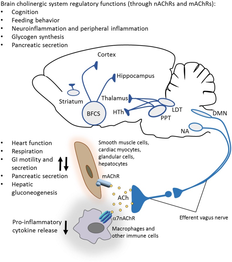
FIGURE 1.
Brain cholinergic system: anatomy and functional control. Important constituents of the brain cholinergic system are the basal forebrain cholinergic system (BFCS) comprised of several nuclei, the mesopontine pedunculopontine and laterodorsal tegmental nuclei (PPT and LDT), and intraneurons in the striatum. Cholinergic neurons provide innervations to different cortex areas, hippocampus, amygdala, olfactory bulb, hypothalamus (HTh), thalamus, and other regions. In addition to its well-documented role in the regulation of cognition, brain cholinergic signaling is implicated in the regulation of appetite and feeding behavior, local brain inflammatory responses (neuroinflammation), hepatic glycogen synthesis, pancreatic secretion, and control of peripheral inflammation (through vagus nerve-mediated mechanisms). Cholinergic neurons in the brainstem dorsal motor nucleus of the vagus (DMN) and nucleus ambiguus (NA) provide axonal projections within preganglionic efferent vagus nerve fibers. These long fibers interact with short postganglionic neurons in proximity or within the innervated organs, including the heart, lungs, gastrointestinal tract, liver, and pancreas. Acetylcholine (ACh) released from these neurons interacts with muscarinic acetylcholine receptors (mAChRs) on targeted cells and regulates several metabolic functions. ACh also regulates (inhibits) the release of pro-inflammatory cytokines and inflammation via alpha 7 nicotinic acetylcholine receptors (α7nAChRs) on immune cells (see the text for more details). (A rodent brain is shown as a substantial part of the information summarized is based on preclinical studies.)

