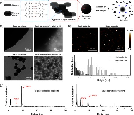
Fig. 1.
Characterization of natural eumelanin models. (a) Schematic illustration of the self-assembled structure of eumelanin and deaggregation process induced by alkaline pH; (b) TEM images of sepia and squid eumelanin before and after exposure to alkaline pH condition in the absence of oxygen for 6 h. (c) AFM analysis of deaggregated subunits from sepia and squid eumelanin. Subunits obtained from deaggregation of sepia and squid eumelanin are deposited on a spinning mica substrate before analysis. (d) HPLC chromatograms of degradation products from sepia and squid eumelanin during alkaline hydrogen peroxide oxidation. PDCA and PTCA appear at 2.6 and 4.6 min, respectively.

