Fig. 2.
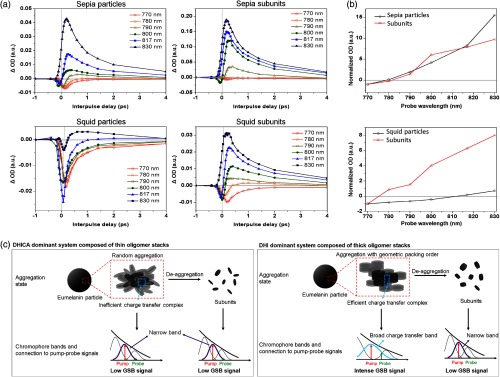
Effect of aggregation on pump-probe signal of eumelanin: (a) 720 nm pump and variable probe signal of eumelanin models and their subunits obtained from deaggregation process under deoxygenated alkaline condition. (b) Time-delayed transient signal at 2 ps as a function of probe wavelength. Signal is normalized at 770 nm. (c) Schematic summary of the proposed relationship between the aggregation-dependent chromophore band of eumelanin and the resulting pump-probe signal. Sepia eumelanin particles, with thin oligomer stacks, do not lead to the efficient edge-to-edge interaction because of low geometric packing preference between oligomer stacks in large-scale aggregation system. Thus, eumelanin particles formed by aggregation of thin oligomer stacks show very similar chromophore bands with their subunits. Because chromophore bandwidth for sepia eumelanin is not broad enough to be in resonance with both pump and probe, GSB is negligible and the whole signal is dominated by ESA. In contrast, squid eumelanin is formed by aggregating the thick oligomer stacks. Because of the high geometric packing preference when aggregated, efficient generation of charge–transfer complexes at the edge of oligomer stacks is possible. Aggregation will result in broad charge–transfer bands that can be resonant with both pump and probe, and thus will lead to intense GSB signal. Because broad charge–transfer bands disappear with deaggregation, GSB signals observed in squid eumelanin particles also diminish with deaggregation, resulting in ESA-dominant signals.

