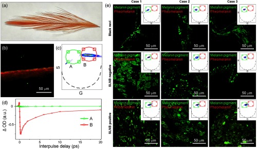
Fig. 6.
Characterization of pheomelanin and its contribution to pigmentation in black nevi, primary melanoma, and metastatic melanoma using 770 nm pump/730 nm probe imaging. (a) Picture of red chicken feather. (b) False-colored images of pheomelanin in red chicken feather derived from (c) the phasor analysis. (d) Pump-probe signal corresponding to regions A and B in the phasor plot (c). Region A corresponds to the signal distribution of eumelanin with different assembly states. Most pump-probe signals of pheomelanin are observed in region B, which clearly differs from that of eumelanin in Fig. 5. (e) Contribution of pheomelanin to pigmentation in black nevi, primary melanoma, and metastatic melanoma. The red component (corresponding to pheomelanin signals) is negligible in all samples.

