Fig. 1.
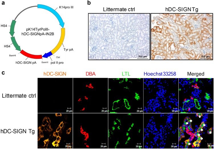
Renal proximal tubular epithelial cells in hDC-SIGN transgenic mice express hDC-SIGN. a Map of the pK14TyrPolII-hDC-SIGNpA-IN2B vector. The human DC-SIGN (hDC-SIGN) gene was cloned under the control of the murine polymerase II large subunit gene promoter, followed by poly(A) tail and two copies of HS4 insulator genes. b, c Kidneys from hDC-SIGN transgenic and littermate control mice were collected after perfusion with PBS. b Paraffin-embedded kidney sections were stained for hDC-SIGN (brown). Slides were counterstained with hematoxylin (blue). Original magnification, ×200. Scale bar = 100 µm. c Cryosections of kidneys were stained for hDC-SIGN (orange), DBA (Dolichos biflorus agglutinin, red), LTL (Lotus tetragonolobus lectin, green), and nuclei (Hoechst 33258, blue). DBA and LTL mark the distal and proximal tubules in the renal cortex, respectively. White arrows point to hDC-SIGN+ cells. Slides were read under a confocal microscope. Original magnification, ×400. Scale bar = 20 µm

