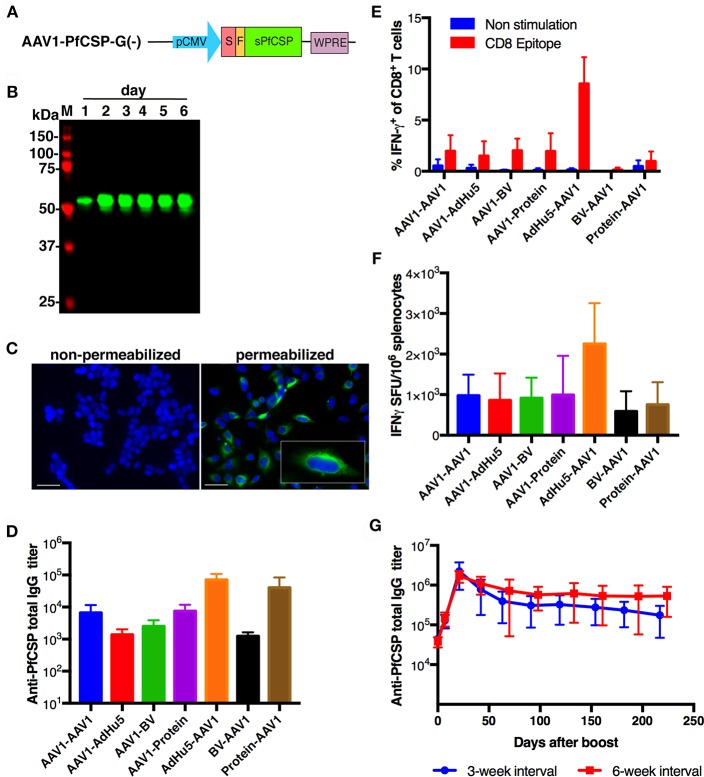
Figure 2.
Functional activity of AAV1-PfCSP-G(–). (A) Construction of AAV1-PfCSP-G(–). Expression of the pfcsp gene cassette was driven by the CMV promoter. S, signal sequence; F, FLAG epitope tag. (B) Expression of PfCSP in HEK293T cells transduced with AAV1-PfCSP-G(–) (MOI = 105), as assessed by immunoblotting with anti-PfCSP mAb 2A10. M, molecular marker. (C) Localization of PfCSP expression in transduced cells. HEK293T cells were transduced with AAV1-PfCSP-G(–) (MOI = 105) as determined by IFA. After 48 h, cells were fixed with methanol (permeabilized) or paraformaldehyde (non-permeabilized) and incubated with Alexa-Fluor-488-conjugated mAb 2A10 (green). Cell nuclei were visualized with 4′,6-diamidino-2-phenylindole (DAPI; blue). Original magnification, ×400. Bars = 50 μm. (D) Anti-PfCSP IgG antibody responses. BALB/c mice (n = 3) were immunized with the indicated regimens at a 3-week interval. Two weeks after boosting, serum samples were collected from each mouse, and their anti-PfCSP IgG titers were determined by ELISA. AdHu5-PfCSP, BV-PfCSP, AAV1-PfCSP-G(–), and rPfCSP protein are shown as AdHu5, BV, AAV1, and protein, respectively. Bars and error bars indicate the means and SD of the values, respectively. (E,F) PfCSP-specific cellular immune responses. BALB/c mice were immunized as described in (D). At 2 weeks post-boost, splenocytes were stimulated with the synthetic PfCSP-specific CD8+ T-cell epitope. (E) An ICS assay was performed on the splenocytes. Percentages of IFN-γ-secreting cells in the CD8+CD4− T-cell population are shown after the subtraction of the percentages of cells stained with an isotype control antibody. (F) An ex vivo ELISpot assay was performed on splenocytes from the same mice. The IFN-γ SFU that reacted with the PfCSP-specific CD8+ T-cell epitope per million splenocytes are shown. (G) Monitoring of anti-PfCSP IgG antibody responses. Groups of BALB/c mice (n = 5) were immunized with an AdHu5-PfCSP -prime and AAV1-PfCSP-G(–)-boost regimen at a 3- or 6-week interval. Serum samples were collected from each mouse 1 day before boost and weekly after boost. Anti-PfCSP IgG titers were determined by ELISA and monitored for 224 days after booster injection.

