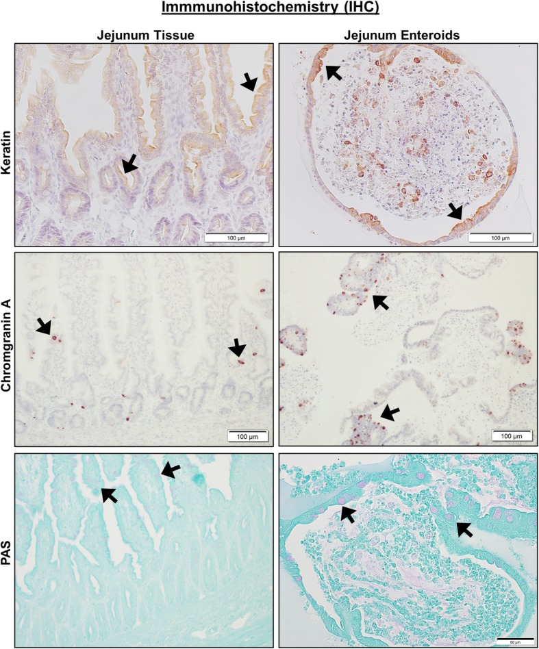
Fig. 4.

Canine jejunal tissues and enteroids both express markers of epithelial cell lineage. Representative immunohistochemistry (IHC) images comparing staining for marker proteins of epithelial cells and their lineage, including Keratin (epithelial cells, upper panels), Chromogranin A (enteroendocrine cells, middle panels), and PAS (goblet cells, lower panels) on both intact whole jejunal tissues and jejunal enteroids. Black arrows indicate representative positive staining for epithelial, enteroendocrine, or goblet cells. Tissue and enteroids were counterstained with Hematoxylin (upper and middle panels) or Alcian Blue (lower panels). Representative images from n = 20 or more enteroids per condition
