Figure 3.
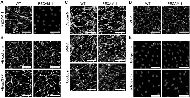
PECAM-1−/− pMBMECs display an intact junctional architecture. Immunofluorescent stainings of WT and PECAM-1−/− pMBMEC monolayers were performed for PECAM-1 (A), VE-cadherin (B), claudin-5 (C), JAM-A (C), occludin (C), and ZO-1 (D). Additionally, the fluorescent signal of VE-CadGFP-expressing pMBMECs is shown (B). Also, stainings for mouse and rabbit isotype controls are presented (E). Cell nuclei were stained with DAPI. Merged grayscale pictures of nucleic signals and the specific antibody stainings are shown. Scale bars, 50 μm.

