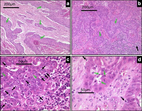
Fig. 3.

Representative histological images of SCCHN tumor sections displaying different levels of immune evasion. a Tumor areas (green arrows) show the absence of any lymphocyte filtration at the rim or within the tumor cell nests. b Lymphocyte infiltrates are seen at the tumor border (black arrows) but are absent within the tumor nests (green arrows). c Despite a high extent of lymphocyte infiltration no signs of tumor cell lysis or apoptosis are visible. d Tumor areas with infiltrating lymphocytes are composed of vital and apoptotic tumor cells (black arrows), indicative of a balance between immune destruction and evasion
