Figure-1.

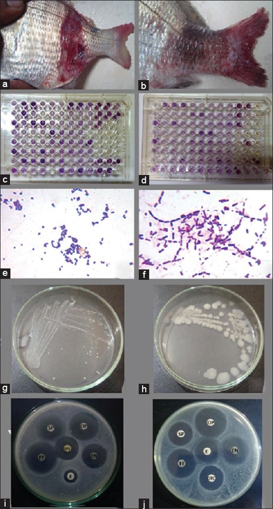
(a and b) Naturally infected white sea bream (Diplodus sargus) showing scale desquamation, skin ulceration even complete loss of skin, and appearance of musculature with congestion and hemorrhage in caudal peduncle together with tail erosion and hemorrhages. (c and d) Biolog GEN III microplate showing the biochemical profile of Staphylococcus epidermidis (c) and Bacillus cereus (d). (e) Gram-stained S. epidermidis appeared as Gram-positive cocci, 0.65-0.91 µm in diameter, present as single, pairs, and clusters. (f) Gram-stained B. cereus appeared as Gram-positive spore-forming long bacilli, 4.97-7.48 µm in length and 1.58-1.64 in width, arranged in short or long chains. (g) S. epidermidis colonies on Tryptic Soy Agar appeared as white pinpoint colonies about 0.2-1 mm in diameter.)h) B. cereus colonies on Tryptic Soy Agar appeared as large white granular colonies with irregular perimeters about 1.5-5 mm in diameter. (i) Antibiogram indicated the sensitivity of B. cereus to oxytetracycline (OTC), ciprofloxacin (CIP), enrofloxacin (ENR), florfenicol (F), and spiramycin (SP), while it resists erythromycin (E). (j) Antibiogram indicated the sensitivity of S. epidermidis to OTC, CIP, ENR, F, SP, and E.
