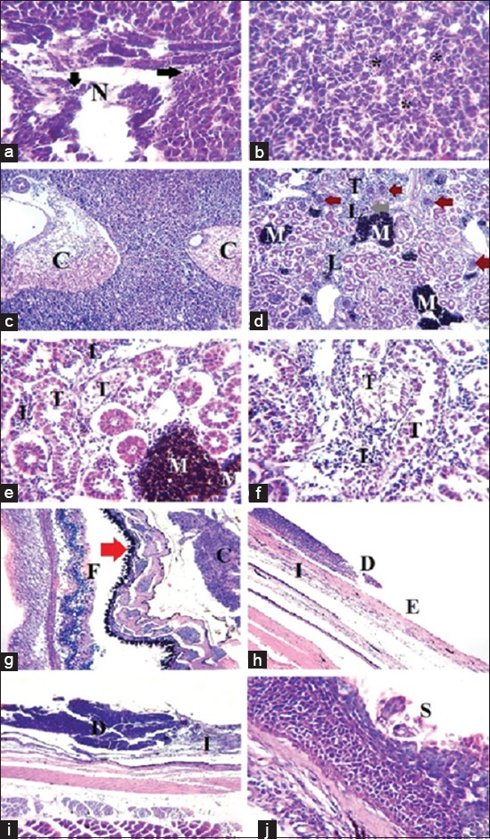
Figure-2.

Histopathological lesions induced by Staphylococcus epidermidis and Bacillus cereus infection in affected white sea bream (Diplodus sargus). (a-c) Hepatopancreas showing loss of normal tissue architecture, presence of necrotic areas (N) with mononuclear cell infiltration (arrow), congested hepatic sinusoids (*) and congested distended blood vessels (C), hematoxylin and eosin (H and E), X=400 in (a and b) and 100 in (c). (d-f) Posterior kidney showing glomerular hypertrophy with narrow Bowman’s space (brown arrow), degenerated shrinkage glomerular taught (gray arrow), detached tubular epithelium (T), interstitial mononuclear cell infiltration (L), melanomacrophage centers activation (M), H and E, X=100 in (d) and 100 in (e and f). (g) Eye with marked separation between retina layers, especially, pigment epithelium layer (red arrow) which also is corrugated and the photoreceptor layer (F), (C) is the choroid body, H and E, X=100. (h-j) Skin showing destruction of epidermis even complete loss (D) with exposure of dermis (E) and presence of leukocytic infiltration (I), in less affected cases loss of the superficial layer of the stratified squamous epithelium (S), H and E, X=100 in (h and i) and 400 in (j).
