


Fig. 8.
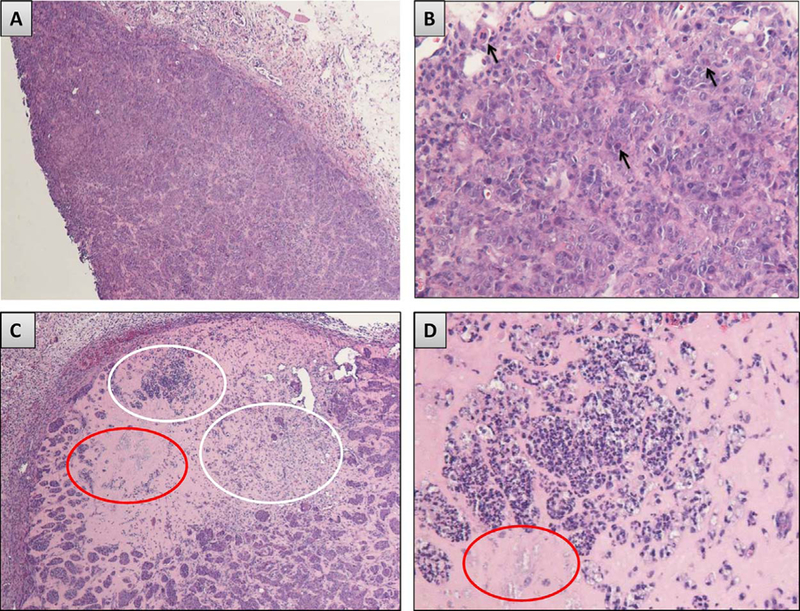
Hematoxylin and eosin (H&E) stained sections from the OSCC tumor cell xenograft studies. A) of OSCC tumor tissue harvested from the control (nontreated) nude mice shows a large aggregation of intact tumor cell nests and cords in a well vascularized tumor stroma. B) Numerous mitotic figures along with scattered apoptotic and dyskeratotic cells are observed in the control group tumor nests. In contrast, C) the 4HPR treated group shows foci of necrosis (white elipse) and residual ghosts of cell nests (red elipse) in residual Matrigel. Appreciably smaller OSCC tumor islands are also visible. D) Higher magnification representation of the necrotic tumor cells and cell island ghosts. Image scale: A, C 40X, B, D 200X.