. 2019 Feb 15;10(14):4004–4014. doi: 10.1039/c9sc00208a

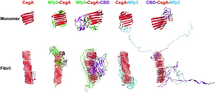
Fig. 6. Comparison of protein monomers and individual fibril structures based upon molecular dynamics simulations. The structures obtained from simulation for the one, two-, and three-component CsgA-fusion protein monomers (top) and fibrils (bottom). Note: for both monomeric and fibrillar states of the constructs, the structures are similar: the CsgA domain always dominate the well-ordered amyloid cores, while disordered Mfp5 or Mfp3 domains as well as CBD are external to the amyloid cores. In particular, the CBD domain in Mfp5-CsgA-CBD retained secondary and tertiary structure similar to the initial folding, while the secondary and tertiary structure of CBD-CsgA-Mfp3 unfolded over the course of simulation. The simulation times for the monomeric and the fibrillar structures were 1 μs and 100 ns, respectively.

Fig. 6