
Figure 7.
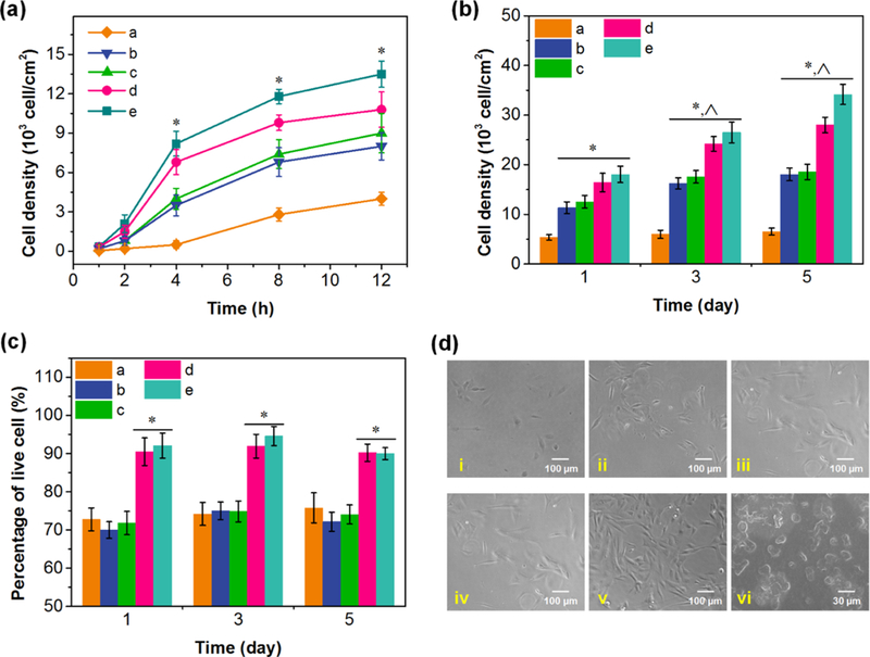
(a) Cell density of osteoblast cells on control substrates, substrates with PL/PG multilayer film, and substrates with PL/PG films incorporated with rhBMP-2 at 1, 2, 4, 8, and 12 h. (b) Proliferation of osteoblast cells and (c) percentage of living cells on various substrates at 1, 3, and 5 days. The samples used in (a)-(c) included (a) quartz slide, (b) L23 film, (c) L5/CG/L8/CL/L8 film, (d, e) L5/CG+BMP/L8/CL/L8 films incubated in rhBMP-2 solution for 5 and 15 min, respectively. (d) Images of osteoblast cell adhesion on samples in (b) and (c) at 24 h. (i-v) Correspond to the samples (a)-(e), respectively. (vi) SEM image of osteoblast cell adhesion. * p < 0.01 compared to quartz, ^ p < 0.01 compared to the same samples cultured at 1 day.