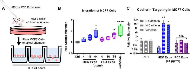
Figure 7.
(A) MCF7 cells were treated with PC3 or HEK exosomes or transfected with 10 nM of hsa-miR-374a-5p. (B) Cellular migration was assessed using a transwell migration assay. The highest dose of PC3 and HEK exosomes induced statistically significant increases in MCF7 migration (p<0.05) as did the positive control miR-374a (p<0.0001). (C) To assess if the increased migration of MCF7 cells upon exposure to HEK exosomes was related to changes in the expression of cadherin genes as predicted, qPCR was performed. HEK exosomes but not PC3 exosomes significantly upregulated the mesenchymal gene markers N-Cadherin and Vimentin (p<0.05, p<0.01) while also displaying a trend toward the reduction of the epithelial marker E-cadherin.

