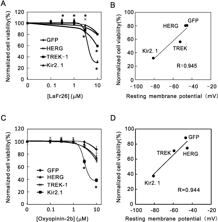
Fig 2. Resting membrane potential-dependency of cytotoxicity of the venom peptides.
(A) LaFr26 was added to the media of 293T cells that stably express GFP, HERG-1, TREK-1, or Kir2.1, and cell viabilities were measured (ANOVA followed Student’s t-test, vs 0 μM, n = 6). (B) A significant correlation was found between cell viabilities at 10 μM and the resting membrane potential measured with whole-cell patch-clamp recordings. (C) Oxyopinin-2b was added to the media of these cells (n = 6). (D) Cell viability at 10 μM again correlated with the resting membrane potentials.

