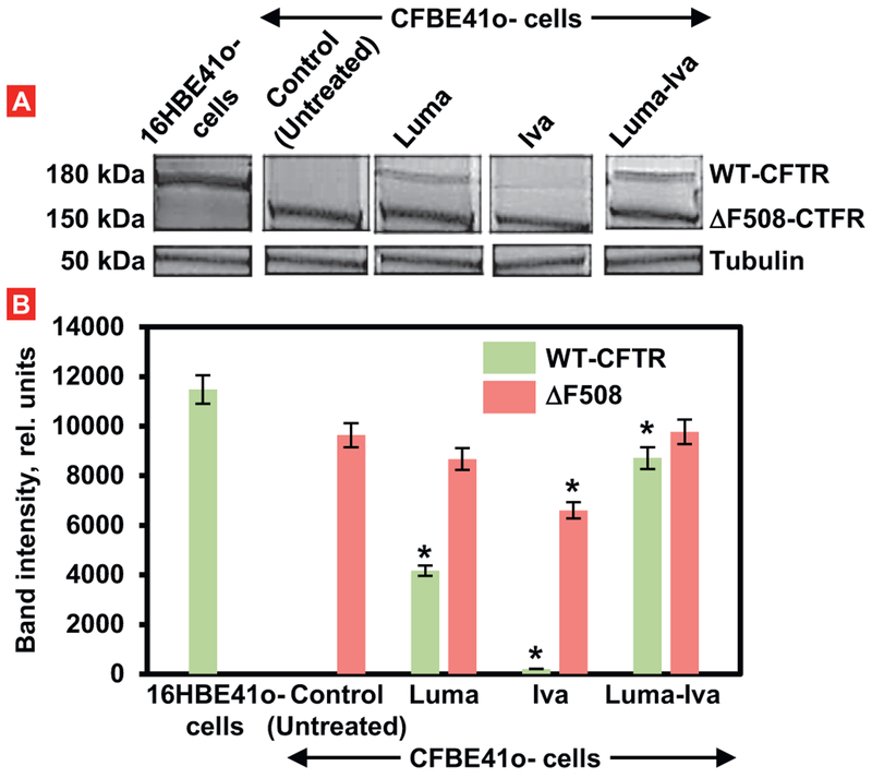
Fig. 4.
Expression of CFTR protein (Western blotting) in healthy (16HBE41o-) and CF (CFBE41o-) human bronchial cells. Two types of proteins: a mature wild type form of CFTR (WT-CFTR, 150 kDa) and mutated (ΔF508-CFTR, 150 kDa) were investigated using tubulin (50 kDa) as an internal standard. CFBE41o-cells were treated for 48 h with 3 μM of free lumacaftor (Luma), ivacaftor (Iva) and their combination. A – typical Western blot image. B – Quantitation of protein expression (Means ± SD are shown). *P < .05 when compared with the expression of corresponding type of protein in untreated CFBE41o- cells.

