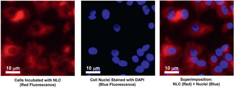
Fig. 5.
Representative images of human bronchial epithelial CFBE41o- cells incubated within 24 h with NLC (red fluorescence). Cell nuclei were stained with nuclear-specific dye (DAPI, blue fluorescence). Superimposition of images allows for detecting of cytoplasmic localization of NLC. (For interpretation of the references to colour in this figure legend, the reader is referred to the web version of this article.)

