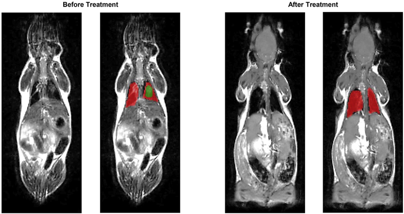
Fig. 6.
Representative magnetic resonance images (MRI) of Tg(FABPCFTR)1Jaw/J homozygote/homozygote bi-transgenic mice with cystic fibrosis before and after treatment. Mice were treated twice per week within four weeks by inhalation with nanostructured lipid carriers containing lumacaftor and ivacaftor. Normal lung tissues are colored in red, while fibrotic tissues – in green. (For interpretation of the references to colour in this figure legend, the reader is referred to the web version of this article.)

