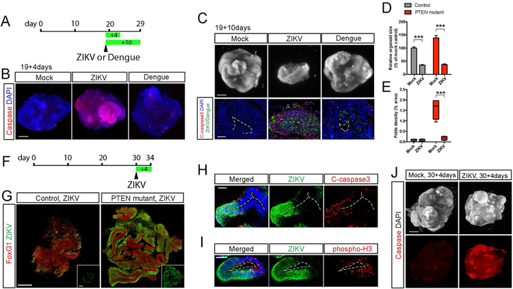
Figure 7. ZIKV infection impairs expansion and folding in human cerebral organoids.
A-B) Schematic diagram and light sheet images of PTEN mutant WIBR3 cerebral organoids at day 23 (19+4 days) showing widespread caspase activity induced by ZIKV but not Dengue virus. Scale bar, 500um.
C) Light sheet images and immuno-staining of mutant WIBR3 organoids at day 29 (19+10 days) shows reduced organoid size and increased apoptosis caused by ZIKV but not Dengue virus. C-caspase 3, cleaved-caspase 3 as detected by immuno-staining. Scale bars, 500um (upper) and 50um (lower).
D-E) Quantitative analysis of organoid at day 29 (19+10 days) shows reduced size (D) and loss of surface folds density (E) upon ZIKV exposure.
F-G) Schematic diagram, and representative images of immuno-staining for ZIKV infected control and mutant WIBR3 organoids at day 34 (30+4 days). Scale bar, 200um.
H-I) Representative images of immuno-staining in mutant WIBR3 organoids show ZIKV infection coincides with elevated apoptosis (cleaved-caspase 3) and reduced proliferation (phosphorylated-H3). Scale bars, 50um.
J) Light sheet images show mutant WIBR3 organoids treated with ZIKV at day 30 displayed widespread apoptosis, as revealed by whole-mount caspase activity staining. Scale bar, 500um.
Results are mean +/− SEM. ***p<0.001.

