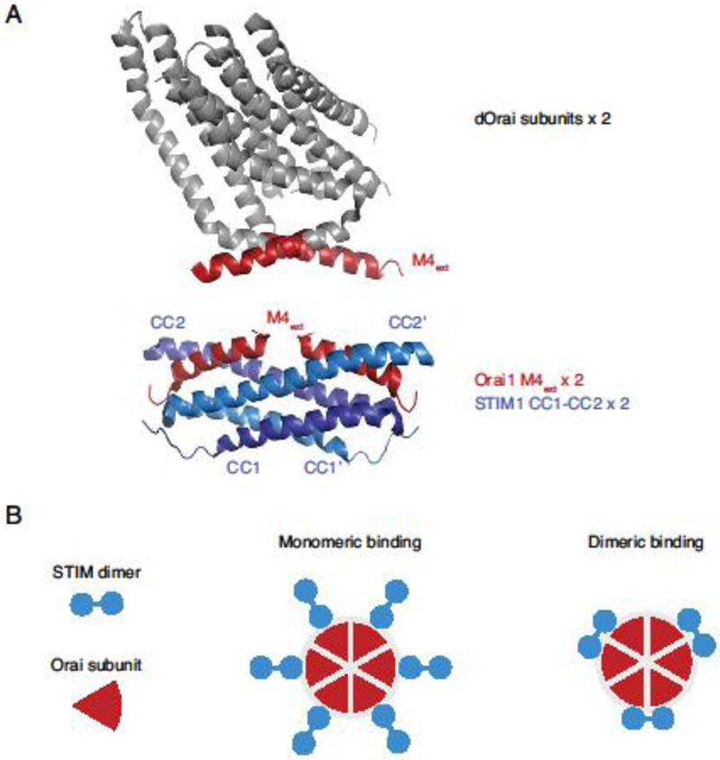
Figure 4. Dimeric and monomeric models of STIM1-Orai1 binding.

A. An example of a dimeric STIM-Orai binding model. Two subunits from the crystal structure of dOrai [51] are shown at top, with the cytoplasmic M4 regions marked in red (4HKR.pdb). The NMR structure below [70] shows two human Orai1 M4 fragments (aa 272-292) bound to a dimer of CC1-CC2 STIM1 fragments (aa 312-383) (2MAK.pdb). B. Schematic views of monomeric and dimeric binding models in which six or three STIM1 dimers are bound to the six Orai1 C termini, respectively.
