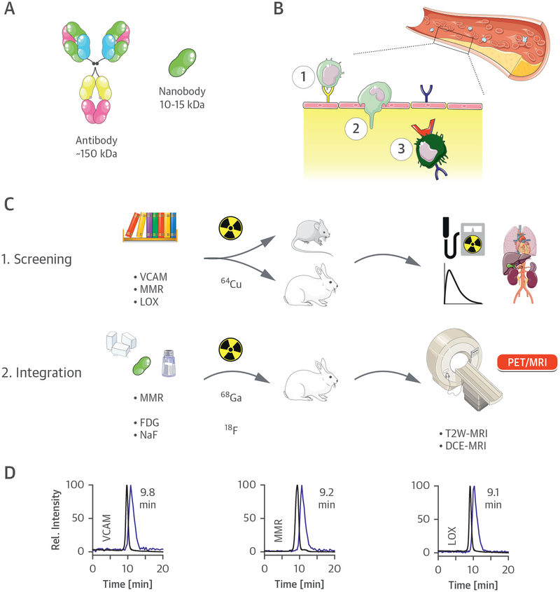
FIGURE 1. Nanobody-Facilitated Atherosclerosis PET Imaging.
(A) Schematic representation of a full-size antibody and a nanobody. (B) Monocyte/macrophage dynamics in atherosclerosis. Endothelial dysfunction is accompanied by the expression of the surface receptor lectin-like oxidized low-density lipoprotein receptor (LOX)-1 (blue) or adhesion molecules like vascular cell adhesion molecule (VCAM)-1 (yellow). Circulating monocytes are recruited to atherosclerotic lesions via interaction with these adhesion molecules (1), leading to infiltration through the endothelium (2). Infiltrated monocytes eventually differentiate into macrophages (3), which may also express LOX-1 and the macrophage mannose receptor (MMR) (red). (C) Study outline. (D) Size exclusion chromatograms of the 3 copper-64 (64Cu) nanobodies, demonstrating coelution of radioactivity (blue trace) with the nonradioactive species (black trace) (λabs = 220 nm). DCE = dynamic contrast enhanced; 18F =fluorine-18; FDG =fluorodeoxyglucose; 68Ga = gallium-68; MRI = magnetic resonance imaging; PET = positron emission tomography; T2W = T2-weighted.

