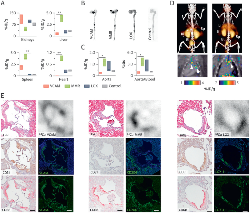
FIGURE 2. Nanobody-Radiotracer Screening in Mice.
(A) Radioactivity distribution in selected tissues in Apoe–/– mice at 3 h post-injection of the corresponding 64Cu-nanobody (n ≥ 3 per nanobody). Autoradiography (B) and radioactivity concentration (C) concentration in aortas of Apoe–/– mice at 3 h post-injection of the corresponding 64Cu-nanobody (n ≥ 3 per nanobody). (D) Representative fused PET/CT images 1 h post-injection of 64Cu-VCAM (left) and 64Cu-MMR (right) in Apoe–/– mice. Arrows indicate enhanced uptake at the aortic arch and root, typical sites of atherosclerotic lesions. (E) Representative images of aortic root sections from Apoe–/– mice with atherosclerosis showing, in the left column, hematoxylin and eosin (H&E) staining (top) and immunohistochemistry for CD31 (endothelial cells) (middle) and CD68 (macrophages) (bottom); in the right column, autoradiography (top) and immunofluorescence for the respective targets of the 3 nanobodies with (middle) and without (bottom) 4,6-diamino-2-phenylindole (DAPI) stain. Bar = 200 μm. *p < 0.05, and **p < 0.01. %ID/g = percentage injected dose per gram of tissue; Bl = bladder; Ki = kidney; Li = liver; Sp = spleen; other abbreviations as in Figure 1.

