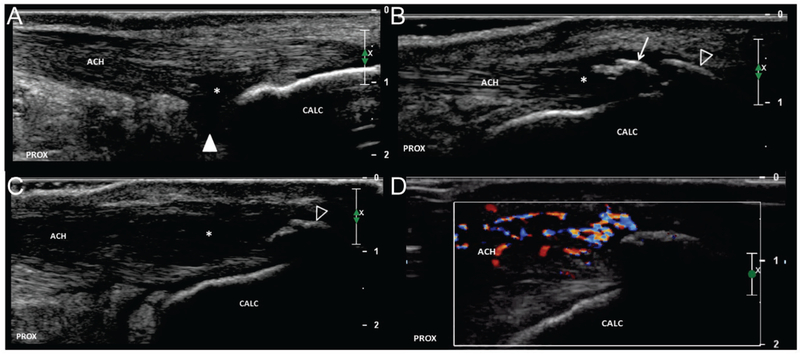
FIGURE 1. Variations in insertional Achilles tendon pathology.
Long axis images of Achilles tendon insertion demonstrating variability in location and extent of pathologic findings. a.) Hypoechoic changes (asterisks) are demonstrated adjacent to the posterosuperior calcaneus (arrow). The boundary with the retro-calcaneal bursa (arrowhead) is ill-defined. Note the relatively normal appearance of the superficial/posterior portion of the tendon. b.) The deep/anterior portion of the tendon is relatively normal; however, changes of tendinosis (asterisks) are appreciated adjacent to an intra-tendinous calcification (arrow). There is minimal posterior acoustic shadowing suggesting “soft” calcification which is amendable to percutaneous debridement. An enthesophyte (open arrowhead) demonstrates dense posterior acoustic shadowing consistent with cortical bone. c.) Hypoechoic changes of tendinosis (asterisks) are more extensive and pronounce. An enthesophyte is present (open arrowhead), but no intra-tendinous calcification is appreciated. d.) Corresponding Color Doppler imaging of figure 1c. There is hyperemia within the superficial/posterior tendon as well as paratenon. ACH = Achilles tendon, CALC = calcaneus, PROX = proximal.

