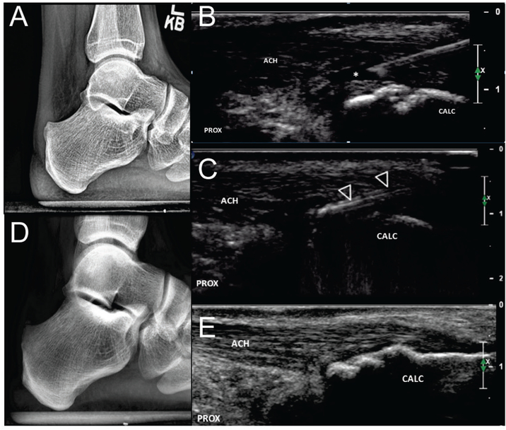
FIGURE 3. Haglund bony debridement.
a.) Pre-procedural X-ray demonstrates a posteriorly projecting bony protuberance at the posterosuperior calcaneus which correlated with location of patient’s maximal pain. b.) Procedural long axis ultrasound image during local anesthesia demonstrates partial thickness tear (asterisks) adjacent to the region of cortical irregularity at the posterosuperior calcaneus (arrowhead). c.) The TX device (open arrowheads) is used to shave down the posteriorly projecting bony protuberance using a layer by layer technique working from superficial to deep. Follow up radiograph at 6 weeks (d.) demonstrates decreased prominence of the previously noted bony protuberance while follow up ultrasound at 3 years (e) is consistent with bony remodeling and complete healing of the debrided partial tendon tear. Patient reports no pain or functional limitation at 3 year follow up. ACH = Achilles tendon, CALC = calcaneus, PROX = proximal.

