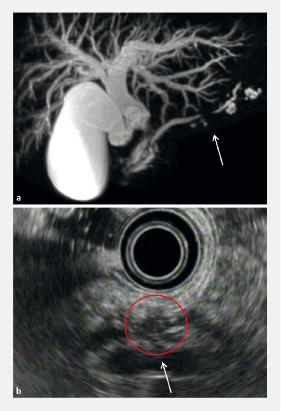
Fig. 2.

Hypoechoic areas in a 66-year-old male with PCIS with cholangiocarcinoma and branch duct IPMN (Case 10). MRCP a and EUS b revealed MPD stricture (arrow). EUS b revealed hypoechoic areas surrounding the MPD stricture, which appeared as a hypoechoic mass (red circle). PCIS, pancreatic carcinoma in situ; MRCP, magnetic resonance cholangiopancreatography; EUS, endoscopic ultrasonography; MPD, main pancreatic duct.
