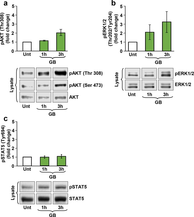
Figure 5.
GB elicited phosphorylation of both Akt and MAPK/ERK kinases in pre-B Nalm-6 cells in a time-dependent manner. Whole cell lysates were subjected to Western blot analysis with the antibodies indicated in material and methods. (a,b) The bar graphs represent densitometry analyses showing a comparable increase in pAkt (Ser 473) and pMAPK/ERK phosphorylation induced after 1 h and 3 h of GB-treated cells (50 µl), respectively (n = 2). As a reference control, lysates from untreated (Unt) pre-B Nalm-6 cells were included, and also, to establish the basal levels of Akt and MAPK/ERK phosphorylation; representative Western blots displaying GB-stimulated pAkt (Ser 473), pAkt (Thr 308) and pERK1/2 phosphorylation are shown in the lower panels. (c) The bar graph represents densitometry analysis showing a comparable increase in STAT5 (Tyr 694) phosphorylation caused after 1 h and 3 h of GB-treated cells (50 µl), respectively (n = 3). As a reference control, lysates from untreated (Unt) pre-B Nalm-6 cells were included; western blots are displaying GB-stimulated STAT5 phosphorylation, as compared with total STAT5 protein are shown in (c), lower panel. The 50 µl of GB correspond to 1.5 ± 0.048 mg/ml of GB lyophilized powder.

