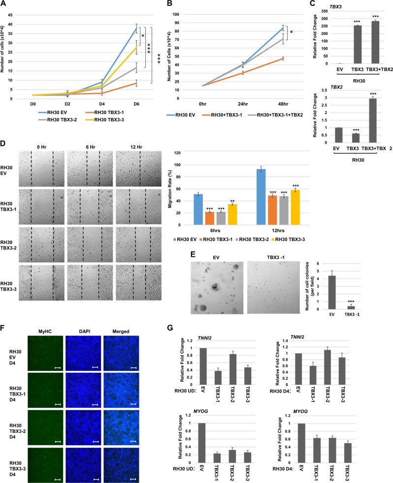
Fig. 3. TBX3 inhibits oncogenic properties but does not promote differentiation of RH30 cells.
a TBX3 inhibits proliferation. Identical number of cells were seeded and cells were counted every 2 days. Error bars, S.E. and *p ≤ 0.05 and ***p ≤ 0.001 versus EV. b TBX2 partially rescues the proliferation defect. The RH30 TBX3-1 cell line was transiently transfected with pEF TBX2 (TBX2) or pEF empty vector (EV) and assayed for proliferation as in (a) except cell counts were performed every 24 h. c mRNA expression of TBX3 and TBX2 in the cells shown in (b) was assayed by qRT-PCR. Error bars, S.E. and ***p ≤ 0.001 versus EV. d TBX3 inhibits migration. Scratch assays were performed on RH30 cells overexpressing TBX3 (TBX3) or pEF (EV). Images were taken at ×100 magnification. Migration rate is quantitated in the right panel. Error bars, S.E. and **p ≤ 0.01, ***p ≤ 0.001 versus EV. e Soft agar assays on RH30 TBX3-1 and EV cell lines. Images were taken at ×100 magnification and the number of colonies was quantified by counting in five random fields. Error bars, S.E. and ***p ≤ 0.001 versus EV. f RH30 overexpressing TBX3 cell lines were assayed by immunofluorescence with myosin heavy chain antibodies (MyHC) following 4 days of differentiation (D4). Images were taken at ×100 and scale bars represent 100 μm. DAPI was used to stain nuclei. g mRNA expression of differentiation specific genes is not upregulated in RH30 cells overexpressing TBX3 in the presence (D4) or absence (UD) of differentiation conditions as assayed by qRT-PCR. Error bars, S.E.

